TrigeminalBalloon Compression
Just different.
The Trigeminal Balloon Compression Cannula Kit was redesigned according to the requirements of the pain treatment procedures related to the trigeminal nerve. Therefore, the new version of the kit made it easier to navigate the balloon catheter, improved the accuracy in the location of the target and, above all, increased safety during the procedure.
Procedure
How is it done?
Usually, the surgery is performed under general anesthesia and the patient is placed in the position with tilted head. The cannula is inserted through a percutaneous puncture under X-ray fluoroscopy. After the cannula is positioned, the balloon catheter is introduced through the cannula and its tip usually protrudes 1 cm beyond the tip of the cannula, about 10 to 22 mm from the target (oval foramen). After that, the non-ionic X-ray contrast agent is injected into the balloon catheter, approximately 0.7 ml (0.5 to 1 ml is usually injected). The puncture is successful when the inflated balloon exibe a pear-like shape.
The compression time of the balloon generally lasts between 1 and 3 minutes. After compression is performed, the contrast agent is removed from the catheter, as well as the balloon catheter and the patient’s cannula.1,2
Myelin
Sheath
One of the most common causes of trigeminal neuralgia is demyelination, or wear of the Myelin Sheath. The Myelin Sheath is a lipoprotein structure that protects nerve cells. This structure that surrounds the trigeminal nerve can be degraded, or degenerated due to a compression of a vein or artery. When this degeneration reaches a more advanced level, the nerve short-circuits, causing pain.3,4
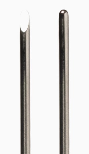
Cannula
New design
Cannula with concave tip and lateral outlet
We reinvented our cannula! In this new design, the internal and external edges are completely rounded. This way it is possible to facilitate the introduction and removal of the catheter without it sliding, breaking or stopping. The surgeon has complete control over the placement of the catheter.
In addition, its new tip ensures smoother advancement and retraction of the cannula, without causing damage to adjacent tissues.
Eliminates cutting, thinning and sliding of the catheter
Smoother feed and retraction
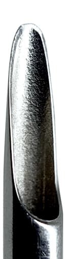
Stylet
As you wish.
The Trigeminal Balloon Compression Cannula Kit comes with two stylets, one with a faceted tip, to facilitate puncture and introduction of the cannula to the desired target and another blunt, allowing fine and precise cannula adjustment without injuring any adjacent tissue in the procedure location.
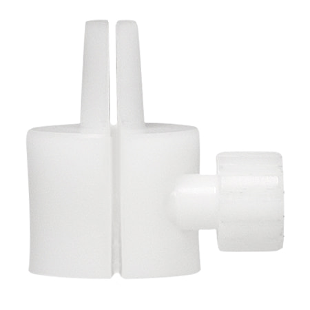
Depth stop
Easier to position the catheter.
Completely new!
The Depth stop is very useful for the correct positioning of the catheter. Usually the tip of the balloon protrudes up to 1 cm beyond the end of the cannula and, with the new design of the Depth stop, it became much more easier.
- Anatomical shape
- Luer connection for coupling to the cannula
- Locking pin with infinite thread (not loose)
- Pin does not lock directly on the balloon (no damage)
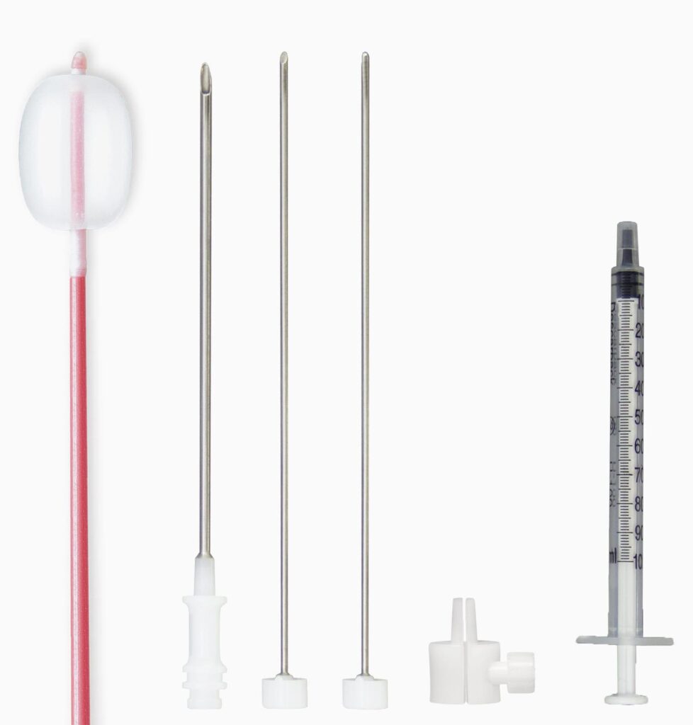
Composition
Trigeminal Balloon Compression Cannula Kit
BMS-D156
- 14G Guide Cannula of 100mm
- Fogarty catheter 4F
- Faceted stylet
- Blunt stylet
- Depth stop
- 1.0ml Syringe